|
||||||||||||||||||||||
|
||||||||||||||||||||||
|
|
||||||||||||||||||||||
|
1.
Introduction
Breast cancer is a leading cause of cancer deaths among women. However
the early detection for cancerous tissues can help reduce the mortality
rates. Until now, the best way for detecting the cancerous tissues is via the
use of mammographic images. The problem with mammography is the large number
of screenings that produces a large number of mammography images, those
images need to be thoroughly examined by radiologists and doctors which is
both financially consuming and time inefficient. Added to that false
detection can cause a high number of unnecessary biopsies. To overcome the
previously mentioned problems a lot of research has been dedicated to
developing computer aided detection applications that can help reduce the
work load and improve the detection rates. This project is an example of the
ongoing research regarding that topic and is conducted in the image and video
systems lab of the Korean advanced institute of technology. 2. Project Goals -
Single-view
mass detection with an improved classification design. -
Improving
mass detection using two views (CC/MLO). -
Micro-calcification
detection from a single-view. -
Integrated
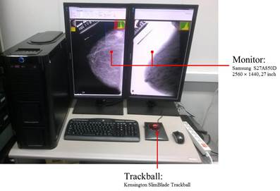
mass and micro-calcification detection. 3. Hardware Configuration
Figure 1 show the demo KAIST-Mammo
CAD system. As the figure shows the demo setup uses two monitors with QHD
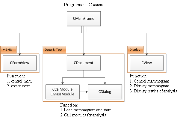
(2550X1440) as well as a trackball. 4. Software Architecture and Functionality 4.1.
Software Architecture:
Figure 2 shows the basic class diagram showing the
basic skeleton for the KAIST-Mammo CAD system. The
system was developed and built using Microsoft Visual C++ 10.0 based on MFC. 4.2. KAIST-Mammo CAD
System Functionality:
Figure 3 show the main pane for the demo KAIST-Mammo CAD System, by adjusting the controllers in this
pane we can perform the following functionalities: -
Single-View
Mass Detection. -
Two-View
Mass Detection. -
Micro-calcification
Detection. -
Combined
Mass and Micro-calcifications Detection. -
Viewer
Aiding Tools. Those functionalities will be further discussed in
the following subsections. 4.2.1. Single-View Mass Detection
Figure 4 shows the configuration for the single-view
mass detection. The demo KAIST-Mammo CAD System
provides the functionality to detect a mass from one of the views (CC or MLO)
in mammograms. The system detects mass areas by marking a blue line around
the detected region. The detected mass regions can be adjusted by adjusting
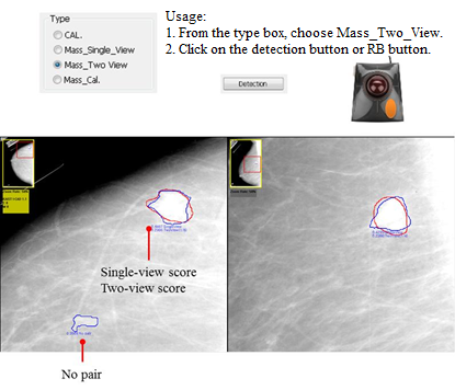
the threshold scroll bar (confidence values). 4.2.2. Two-View Mass Detection
Figure 5 shows the configuration for the two-view
mass detection. The demo KAIST-Mammo CAD System
provides the functionality to detect a mass from both of the mammography
views (CC and MLO) for the same breast laterality. The system detects pairs
detected ROIs one from each view and classify it as a mass accordingly. 4.2.3. Microcalcification Detection
Figure 6 shows the configuration for the
micro-calcification detection. The demo KAIST-Mammo
CAD System provides the functionality to detect micro-calcification from one
mammography view (CC or MLO). The system detects micro-calcification cluster
regions by marks them with blue triangles. 4.2.4. Combined Mass and Micro-calcification
Detection
Figure 7 shows the configuration for the combined
mass and micro-calcification detection. The demo KAIST-Mammo
CAD System provides the functionality to detect both masses and
micro-calcification clusters from a single mammography view (CC or MLO). In
this configuration the system gives higher weights for mass regions with
micro-calcifications inside them. 4.2.5. Viewer Aiding Tools The following tools were also developed in order to
help the radiologist while viewing the mammograms. -
Zoom in/out,
magnification and moving. -
Intensity change
and inversion. -
Control the
size of ROIs(mass, calcification) |
||||||||||||||||||||||
|
|
||||||||||||||||||||||


|
|
![]()
- Contact Person: Prof. Yong Man Ro (ymro@kaist.ac.kr)