|
1.
Introduction
l
Recent
clinical studies have shown that computer-aided detection (CAD) systems could
play an important role in increasing the breast cancer
detection rate in mammography.
l
The
detection of breast masses on mammograms (especially, in case of dense
breast tissue) is far from being mature stage as masses generally have similar
intensity with their surroundings.
l
Mass
detection in dense mammograms is critical because several studies have
shown that tissue types are related with breast cancer risks.
l
We
propose a novel mammogram enhancement for improving mass detection accuracy.
The proposed enhancement method based on two observations on masses.
l
Masses
are hyper-dense or uniform density with respect to their background.
l
The
core parts of masses have high intensity while intensities are decreased
as the distance to core parts is increased.
2.
Image enhancement
2.1.
Purpose
l
Enhancing
regions which have aforementioned both mass characteristics
simultaneously.
2.2. Method
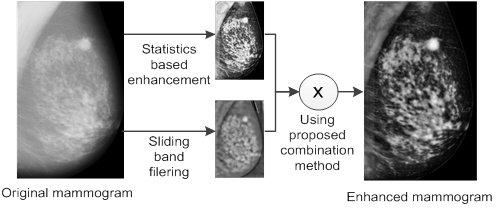
Fig. 1.
Block diagram of the proposed enhancement method
l
Enhancing
aforementioned mass characteristics separately, then combining as
follows:
l
ISTAT,
ISBF
: Filter responses of the statistics based
enhancement and the sliding band filtering, whose values are normalized
to [0, 1].
l
α1,
α2
: Weights of each filter response.
l
ⓧ
: Pixel-wise multiplication operator.
l
Statistics
based enhancement
n
Enhancing
bright and smooth regions and suppressing backgrounds.
l
Sliding
band filtering
n
Enhancing
the regions where their surrounding gradients are converging regardless
of the contrast of the regions.
3.
Experiments
3.1.
Experimental setup
l
89
mammograms collected from mini-MIAS DB were used.
l
Contour-based
detection algorithm was adopted for the detection.
l The correct detection was recorded for a segmented region
when the region included the centroid of a mass.
3.2.
Effectiveness of the proposed enhancement
l
The
proposed enhancement increased the overall mass detection accuracy. In
particular, the detection accuracy is significantly increased in dense parenchyme.
Table 1. Mass detection sensitivity of all abnormalities
|
Mass detection method
|
Mass detection sensitivity
|
|
Contour-based detection
without the proposed enhancement
|
78.3%
|
|
Contour-based detection
with the proposed enhancement
|
92.4%
|
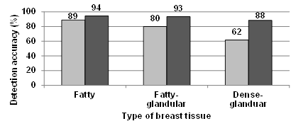
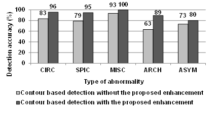
Fig. 2. Mass detection accuracy, with respect to the three
different types of breast tissue (up)
and to the five different types of abnormality (down)
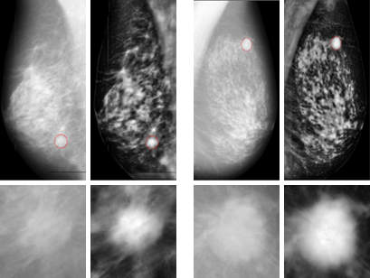
Fig. 3. Examples of the original mammograms and mammograms
enhanced
by the proposed method (breast tissues of both mammograms are dense-glandular)
4. Conclusions
l
The proposed
enhancement is designed for increasing contrast between mass and
backgrounds.
l
The
results demonstrate that the proposed enhancement can significantly
improves the detection accuracy of the mass detection, especially
detection accuracy of masses in dense mammogram.
|

