|
1. Osteoporosis
|
|
%20▒%20▒%20▒_files/dot1.gif)
|
Over the 50 percents of women who are older than 45
years have osteoporosis
|
|
|
People hardly recognize this disease by themselves
|
|
|
Measuring BMD(Bone Mineral Density) have been done
widely to detect osteoporosis in the early stage
|
|
|
To measure BMD correctly, it is important to segment
bone area and measure de-noised biomedical signal
|
|
|
|
|
2. Mammography
|
|
|
X-ray image is obtained from absorption or scattering
effect of photons that is emitted from X-ray energy source based on
characteristic or distribution of materials that consist of the object
|
|
|
DEXA is the method based on two different X-ray energy
levels obtained high energy and low energy level
|
|
DEXA is an effective tool to minimize anatomic noise
which is main problem in medical X-ray analysis by two district X-ray
energy images
|
|
|
|
|
Fig 1. Principle of DEXA Image
|
|
3. Body DEXA Image Decomposition based on Attenuation
Coefficient
|
|
|
Materials composing human body roughly divide into bone
and soft tissue region which have the unique attenuation coefficient
|
|
|
Attenuation in permeation of X-ray can be represented by
combination of bone and soft tissue
|
|
|
|
|
|
Fig 2. DEXA Image Decomposition
|
|
4. DEXA Image Noise Modeling and Reduction
|
|
A) DEXA image noise modeling
|
|
DEXA image noise modeling based on the analysis of
characteristics of system input/output noise
|
|
|
DEXA image noise model
|
|
1)Source noise
characteristics of DEXA image

2)Detector noise
characteristics of DEXA image
|
B) DEXA Image Denoising
|
|
Noise exists multiplicative and linear additive noise in DEXA image
|
|
|
Denoising method consists of two : Linear noise
reduction and multiplicative noise reduction using logarithm and
wavelet shrinkage method
|

Fig 3. DEXA Image Noise Reduction Method
|
|
Example of Denoised DEXA Image
|
|
|
5. Segmentation for distal radius bone
|
|
|
Active
Shape Model (ASM) is effective technique in bone segmentation
|
|
|
|
Chamfer
matching find the initial
rotation, scale, and translation of ASM
segmentation
|
|
|
|
Using
DXA decomposition, we eliminate effect of the soft tissue
|
|
|
|
Weight
based on decomposition provide the solution for multi edge problem for
ASM in distal radius bone
|
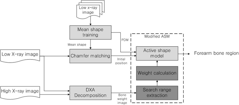
Fig 4. ASM segmentation system in multi edge environment
|
|
|
Segmemtation Result for example image
|
|
|
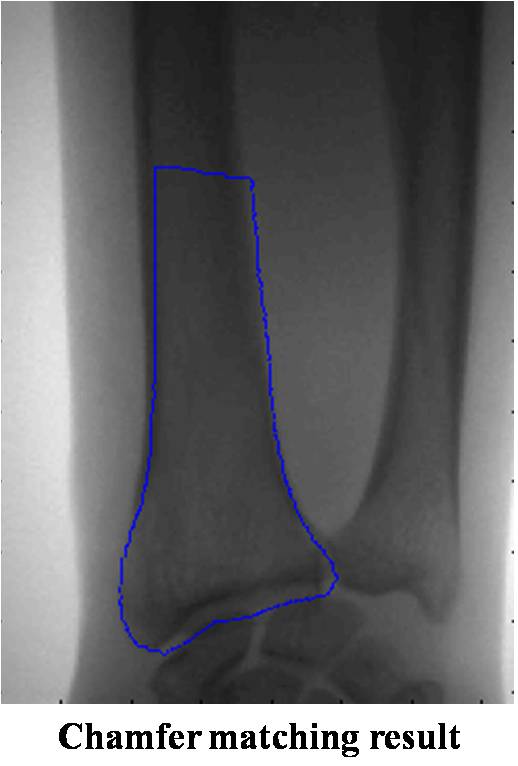 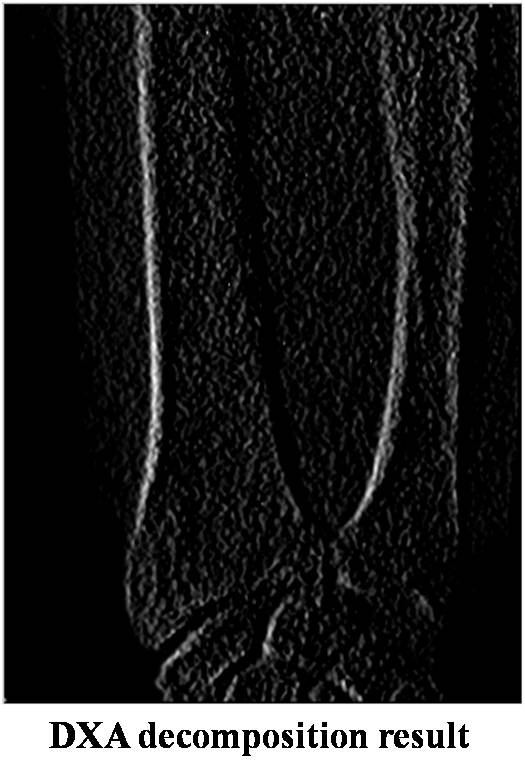 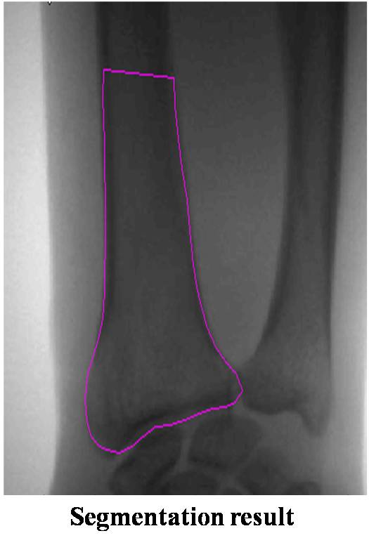
Fig 6. Segmentation result
|
|
|
| |
%20▒%20▒%20▒_files/project_title.gif)